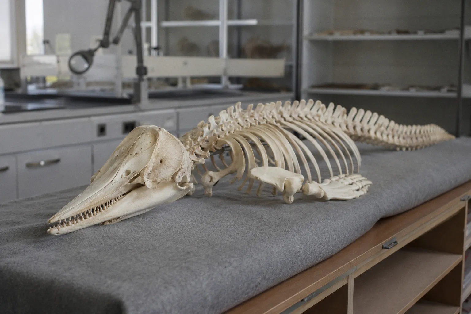
Skeleton of the world’s rarest marine mammal preserved by digital imaging
Resolving skeletal structures at tolerances below the width of a human hair requires a hybrid imaging pipeline. The methodology combines macro-photography, medical-grade computed tomography (CT), and micro-CT scanning.
Multi-Modal Scan Acquisition and Spatial Resolution
The data acquisition process relied on dual-tier CT scanning to capture both macro-scale geometry and micro-scale bone density. First, hospital-grade CT scanners captured the overall skeletal framework of the vaquita, a small porpoise native to Mexico's northern Gulf of California that is currently limited to an estimated population of seven to 10 individuals.
To resolve fine structural details, the team integrated microscopic CT imaging. This modality operates at spatial resolutions capable of capturing features smaller than a human hair. By combining these scans with high-resolution photography, the imaging pipeline captured the surface texture and internal geometry without subjecting the 1966 physical specimen to mechanical stress or environmental degradation.
Dataset Assembly and Reconstruction Methodology
Post-acquisition processing required the alignment and assembly of thousands of individual scan slices. These slices were compiled into a unified three-dimensional model representing every bone in the specimen's skeletal structure.
* Capture Target: Complete female vaquita skeleton (1966 origin).
* Sensing Modalities: Hospital-grade CT, micro-CT, high-resolution photography.
* Spatial Tolerance: Sub-human-hair width.
* Data Output: Multi-thousand slice volumetric reconstruction.
* Distribution Metric: Open-access digital dataset.
This structured workflow eliminates the typical degradation risks associated with physical mold-making or repeated manual handling of fragile biological specimens. The resulting digital assets allow for the production of physical replicas via 3D printing for educational and museum deployments.
Digital Archiving Frameworks and Industry Standards
This project aligns with broader institutional digitization initiatives, including the oVert project in the United States and Ozboneviz in Australia. These frameworks leverage advanced imaging pipelines to convert restricted-access museum collections into open-source digital assets.
For technical specialists, the standardization of these multi-modal imaging pipelines provides a repeatable blueprint for archiving rare physical media, historical artifacts, and delicate structural components. The resulting datasets allow global researchers to conduct volumetric and structural analyses without geographic or physical access limitations.
Verdict
Strictly evaluated, the hybrid CT and micro-CT imaging pipeline delivers the required spatial resolution to preserve fragile physical specimens digitally, though the processing overhead of assembling thousands of high-resolution slices remains a significant computational bottleneck.